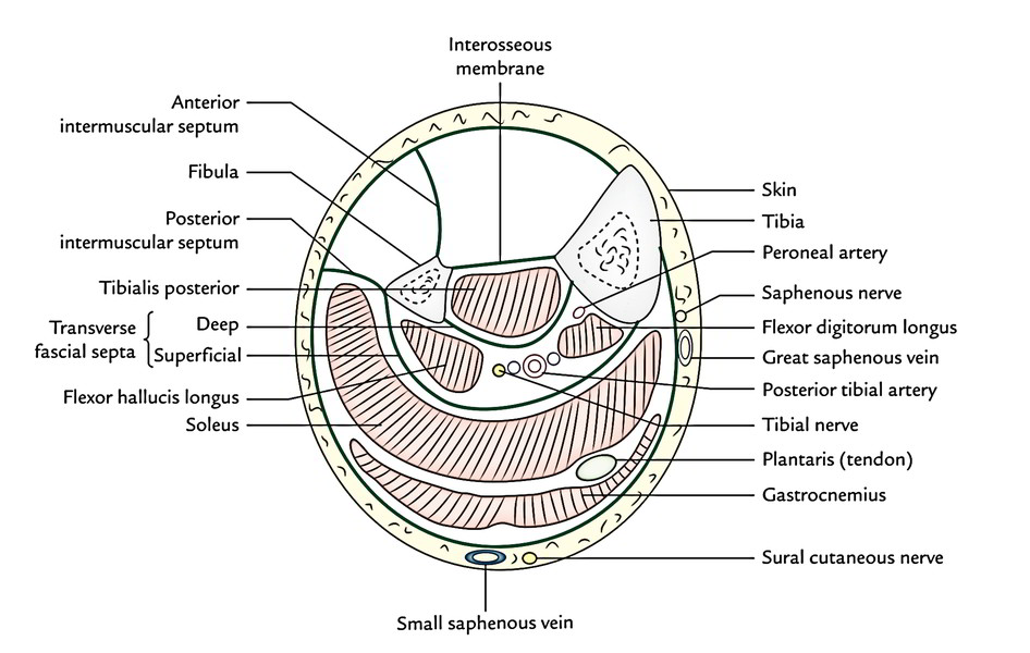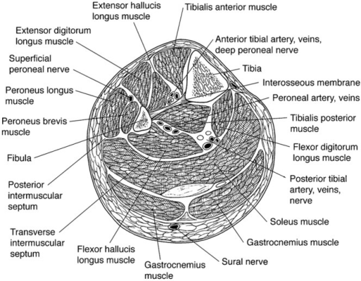
21-24 ) passes into the plantar surface of the foot to form part of the PLANTAR ARCH. It goes up the back of the leg, and passes through the deep fascia near. The posterior muscle group is the smallest group, occupying the posterior compartment of the thigh. The short saphenous vein runs up between the calcaneal tendon and the lateral malleolus. The anterior compartment of the leg is supplied by the deep fibular nerve (deep peroneal nerve), a branch of the common fibular nerve.The nerve contains axons from the L4, L5, and S1 spinal nerves. These muscles are also called the adductors of the thigh. The compartment contains muscles that are dorsiflexors and participate in inversion and eversion of the foot. The posterior tibial and peroneal vessels and the tibial nerve lie between the deep muscles and deep septum at the level of the calf. It includes the pectineus, adductor magnus, adductor minimus, adductor longus, adductor brevis, and gracilis. The superficial compartment containing the gastrocnemius and soleus muscles is separated from the deep posterior compartment containing the plantar flexors by the deep septum spanning from the tibia to the fibula.

The first dorsal metatarsal artery runs distally in the first interspace and divides into dorsal digital arteries to the medial and lateral sides of the big toe and the medial side of the second toe. This video describes the anatomy of the posterior compartment of the leg as regard posterior tibial nerve, posterior tibial artery, peroneal artery and fle. The medial group occupies, you guessed it, the medial compartment of the thigh. 21-24 ) to the first interspace between the first and second metatarsal arises where the deep plantar artery pierces this first interspace to reach the plantar surface of the foot. The lateral most dorsal metatarsal artery also gives off a lateral branch to the lateral side of the little toe. Each dorsal metatarsal artery divides into DORSAL DIGITAL ARTERIES that run along the adjacent sides of the 2-5 toes. It gives off three DORSAL METATARSAL ARTERIES that run in the spaces between the 2-3, 3-4, and 4-5 metatarsals.
COMPARTMENTS OF LEG VESSELS SKIN
The fascia surrounding the calf and thigh muscles separates two compartments: the superficial compartment, consisting of all tissues between the skin and the.

Compartments are located in the arms, hands, feet, and legs. What are the arteries of the posterior compartment Posterior tibial and fibular vessels.

As previously mentioned, they are dorsiflexors. Fascia does not easily expand, so if swelling causes pressure to build within a compartment, you need treatment to alleviate the pressure. Deep fibular nerve, dorsiflexors of the foot and toes. The muscles found in the anterior compartment of the leg are: the tibialis anterior, extensor hallucis longus, extensor digitorum longus and fibularis tertius muscle. The ARCUATE ARTERY curves laterally off the do rsalis pedis and runs along the metatarsal bases, deep to the extensor digitorum brevis (Fig. Thorough knowledge of the fascial compartments of the leg is a prerequisite of understanding the relationship between superficial and deep veins. Muscle compartments are groups of muscles, nerves, and blood vessels wrapped in thick sheets of connective tissue called fascia.


 0 kommentar(er)
